
Mouse Secondary Antibodies

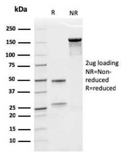
Biotium Nucleophosmin (Acute Myeloid Leukemia Marker)(rNPM1/1901), 0.2mg/mL
Recognizes a 33 kDa glycoprotein, identified as Nucleophosmin (NPM). It is predominantly localized in the nucleus of cells in most tissues. NPM is involved in ribosomal assembly and rRNA transport. It is an abundant protein that is highly phosphorylated by Cdc2 kinase during mitosis. This phosphoprotein moves between the nucleus and the cytoplasm. It is thought to be involved in several processes including regulation of the ARF/p53 pathway. A number of genes are fusion partners, in particular the anaplastic lymphoma kinase gene on chromosome 2. Mutations in exon 12 affecting the C-terminus of the protein are associated with an aberrant cytoplasmic location. Mutations in this gene are associated with acute myeloid leukemia. The antibody may be a useful aid for classification of acute myeloid leukemia. Primary antibodies are available purified, or with a selection of fluorescent CF Dyes and other labels. CF Dyes offer exceptional brightness and photostability. Note: Conjugat

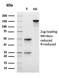
Biotium Erythropoietin (EPO) (Marker of Placentation Disorders)(rEPO/1367), Biotin conjugate, 0.1mg/mL
Recognizes a protein of about 37 kDa, which is identified as Erythropoietin (EPO). Erythropoietin is a secreted, glycosylated cytokine hormone composed of four alpha helical bundles. It is the primary factor responsible for regulating erythropoiesis during steady-state conditions and in response to blood loss and hemorrhage in the adult organism. Erythropoietin is synthesized by the kidney and stimulates the proliferation and maturation of bone marrow erythroid precursor cells. The protein is found in the plasma and regulates red cell production by promoting erythroid differentiation and initiating hemoglobin synthesis. Primary antibodies are available purified, or with a selection of fluorescent CF Dyes and other labels. CF Dyes offer exceptional brightness and photostability. Note: Conjugates of blue fluorescent dyes like CF405S and CF405M are not recommended for detecting low abundance targets, because blue dyes have lower fluorescence and can give higher non-specific
Biotium Lymphocyte Specific Protein 1 (LSP1) / pp52(LSP1/3042), CF488A conjugate, 0.1mg/mL
LSP1 is a hematopoietic-expressed gene that encodes an F-actin-binding, leukocyte-specific (including B and T lymphocytes, granulocytes and macrophages), phosphoprotein. However, mRNA splice variants that do not encode the lympho-specific protein are expressed from this gene in nonlymphoid cell lines as well (myocytes, stromal cells and fibroblasts), suggesting pp52 has a divergent role in signal transduction. The pp52 (LSP1) locus maps to human chromosome 11p15. 5, which is implicated in tumor-related chromosomal translocations found in chronic lymphocytic leukemia. The pp52 promoter contains key elements that control transcriptional activity including an initiator specifying the unique 5' terminus of pp52 mRNA, tandem pairs of Ets and SP1 motifs, and a single C/EBP motif. LSP1 binds the cytoskeleton and has been implicated in affecting cytoskeletal remodeling in a variety of leukocyte functions, including cell motility and chemotaxis. Primary antibodies are available purifi
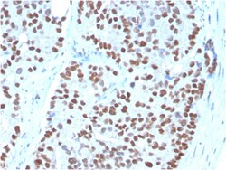
Biotium NKX3.1 (Metastatic Prostate Adenocarcinoma Marker)(NKX3.1/3348), 1mg/mL
NKX3. 1 is a prostate specific gene encoding a transcription factor that plays an important role in normal prostate development and carcinogenesis. It is a prostatic tumor suppressor gene located on chromosome 8p21. 2, which frequently undergoes a loss of heterozygosity. NKX3. 1 expression is highly restricted in prostate epithelial cells and therefore can be used as a diagnostic biomarker for prostate cancer and other metastatic lesions of prostatic origin. Furthermore, NKX3. 1 shows better sensitivity than Prostate Specific Antigen (PSA) for identifying metastatic prostatic adenocarcinoma. This suggests that immunohistochemical staining of NKX3. 1, along with other prostate-restricted markers, may be valuable for the definitive determination of prostatic origin in poorly differentiated metastatic carcinomas. Primary antibodies are available purified, or with a selection of fluorescent CF Dyes and other labels. CF Dyes offer exceptional brightness and photostability. Note
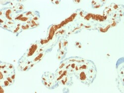
Biotium Spectrin Alpha 1 (Erythrocyte Marker) (SPTA1/1832), CF568 conjugate, 0.1mg/mL
Spectrin is an actin crosslinking and molecular scaffold protein that links the plasma membrane to the actin cytoskeleton, and functions in the determination of cell shape, arrangement of transmembrane proteins, and organization of organelles. Spectrins function as membrane organizers and stabilizers, composed of non-homologous α andβ chains, which aggregate side-to-side in an anti-parallel fashion to form dimers, tetramers and higher polymers. Spectrin α I and spectrinβ I are present in erythrocytes, whereas spectrin α II (also designated fodrin α) and spectrinβ II (also designated fodrinβ) are present in other somatic cells. The spectrin tetramers in erythrocytes act as barriers to lateral diffusion, but spectrin dimers seem to lack this function.Primary antibodies are available purified, or with a selection of fluorescent CF Dyes and other labels. CF Dyes offer exceptional brightness and photostability. Note: Conjugates of blu
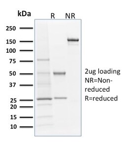
Biotium Langerin / CD207 (Marker of Langerhans Cells)(LGRN/1821), Biotin conjugate, 0.1mg/mL
Langerhans cells (LCs) are a subset of immature dendritic cells (DCs) that specifically localize in the epidermis and other mucosal epithelia. Epidermal LCs possess strong immuno-stimulatory capacity and play a central role in the initiation and regulation of immune responses. Langerin (CD207) is a Ca2 -dependent, C-type lectin domain containing, type II transmembrane protein that induces epidermal LCs to differentiate into Birbeck granules (BG). BGs are organelles with superimposing and zippering membranes that influence proper class I type antigen presentation to the circulating T cells. Human spleen, lymph node, thymus, liver, lung and heart express Langerin protein. Primary antibodies are available purified, or with a selection of fluorescent CF Dyes and other labels. CF Dyes offer exceptional brightness and photostability. Note: Conjugates of blue fluorescent dyes like CF405S and CF405M are not recommended for detecting low abundance targets, because blue dyes have
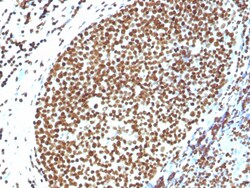
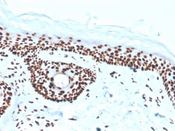
Biotium Histone H1 (Pan Nuclear Marker)(N/A), 1mg/mL
Eukaryotic histones are basic and water-soluble nuclear proteins that form hetero-octameric nucleosome particles by wrapping 146 base pairs of DNA in a left-handed super-helical turn sequentially to form chromosomal fiber. Two molecules of each of the four core histones (H2A, H2B, H3, and H4) form the octamer; formed of two H2A-H2B dimers and two H3-H4 dimers, forming two nearly symmetrical halves by tertiary structure. Over 80% of nucleosomes contain the linker Histone H1, derived from an intronless gene that interacts with linker DNA between nucleosomes and mediates compaction into higher order chromatin. Histones are subject to posttranslational modification by enzymes primarily on their N-terminal tails, but also in their globular domains. Such modifications include methylation, citrullination, acetylation, phosphorylation, sumoylation, ubiquitination and ADP-ribosylation. Primary antibodies are available purified, or with a selection of fluorescent CF Dyes and other lab

Biotium CD10 (Membrane Metalloendopeptidase) (MME/1892), CF568 conjugate, 0.1mg/mL
CD10 (also known as Common Acute Lymphocytic Leukemia Antigen (CALLA)) is a type II transmembrane protein. It is a cell surface enzyme with neutral metalloendopeptidase activity, which inactivates a variety of biologically active peptides. CD10 is expressed on the cells of lymphoblastic, Burkitt's, and follicular germinal center lymphomas, and on cells from patients with chronic myelocytic leukemia (CML). It is also expressed on the surface of normal early lymphoid progenitor cells, immature B cells within adult bone marrow and germinal center B cells within lymphoid tissue.Primary antibodies are available purified, or with a selection of fluorescent CF Dyes and other labels. CF Dyes offer exceptional brightness and photostability. Note: Conjugates of blue fluorescent dyes like CF405S and CF405M are not recommended for detecting low abundance targets, because blue dyes have lower fluorescence and can give higher non-specific background than other dye colors.
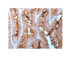
Biotium Carcinoembryonic Antigen (CEA) / CD66 (C66/1983R), 0.2mg/mL
This antibody recognizes proteins of 80-200 kDa, identified as different members of CEA family. CEA is synthesized during development in the fetal gut and is re-expressed in increased amounts in intestinal carcinomas and several other tumors. This MAb does not react with nonspecific cross-reacting antigen (NCA) and with human polymorphonuclear leucocytes. It shows no reaction with a variety of normal tissues and is suitable for staining of formalin/paraffin tissues. CEA is not found in benign glands, stroma, or malignant prostatic cells. Antibody to CEA is useful in detecting early foci of gastric carcinoma and in distinguishing pulmonary adenocarcinomas (60-70% are CEA ) from pleural mesotheliomas (rarely or weakly CEA ). Anti-CEA positivity is seen in adenocarcinomas from the lung, colon, stomach, esophagus, pancreas, gallbadder, urachus, salivary gland, ovary, and endocervix.Primary antibodies are available purified, or with a selection of fluorescent CF Dyes and other la
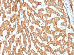
Biotium Prohibitin (Mitochondrial Marker)(PHB/1882), 1mg/mL
Recognizes a protein of 30 kDa which is identified as Prohibitin, an evolutionarily conserved protein with homologues found in yeast to man. It is located in the inner membrane of mitochondria. Although prohibitin mRNA and protein expression occurs throughout the cell cycle, maximum levels are detected during the G1/S phase transition and minimum levels are seen in S phase and the G2/mitosis boundary. Prohibitin is located exclusively in the mitochondria with the highest concentration on the inner membrane. Prohibitin is an ideal mitochondrial marker. It shows antiproliferative activity and has been proposed to play a role in normal cell cycle regulation, replicative senescence, cellular immortalization, and tumor suppression. Primary antibodies are available purified, or with a selection of fluorescent CF Dyes and other labels. CF Dyes offer exceptional brightness and photostability. Note: Conjugates of blue fluorescent dyes like CF405S and CF405M are not recommended fo
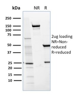
Biotium CD36 (Platelet & Microvessel Marker)(GPIIIb/1654), CF740 conjugate, 0.1mg/mL
Recognizes a protein of 80 kDa-90 kDa, identified as CD36. It is expressed on platelets, monocytes and macrophages, microvascular endothelial cells, erythrocyte precursors, mammary epithelial cells, and some macrophage derived dendritic cells. CD36 acts as a receptor for thrombospondin (TSP), collagen types I, IV and V, P. falciparum malaria-infected erythrocytes, and sickle erythrocytes. It also functions as a scavenger receptor, mediating macrophage uptake of oxidized low-density lipoprotein (LDL) and recognition of apoptotic polymorphonuclear leukocytes (PMN). CD36 plays a role in platelet aggregation, macrophage foam cell development, inflammation, and the tissue ischemia observed in sickle cell disease and cerebral malaria. Note that 1-4% of Japanese and East Asia population lack CD36. Primary antibodies are available purified, or with a selection of fluorescent CF Dyes and other labels. CF Dyes offer exceptional brightness and photostability. Note: Conjugates of blue
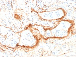
Biotium Elastin (ELN) (Marker of Arterial Stiffness and Atherosclerosis) (ELN/1981), CF740 conjugate, 0.1mg/mL
Elastin is a polymeric protein and is the main component of the extracellular matrix of arteries. It is synthesized and secreted as a soluble, single-chain protein (tropoelastin), which undergoes a number of post-translational modifications prior to its organization into an elastic fiber in the extracellular space. Elastin performs a regulatory function during arterial development by controlling proliferation of smooth muscle and stabilizing arterial structure. It imparts elasticity to the connective tissue. During aging, the elasticity of connective tissue is reduced because of the cross-linking of collagenous fibers with elastin. The abnormal accumulation of elastic tissue in blood vessels is found in atherosclerosis and hypertension.Primary antibodies are available purified, or with a selection of fluorescent CF Dyes and other labels. CF Dyes offer exceptional brightness and photostability. Note: Conjugates of blue fluorescent dyes like CF405S and CF405M are not recom
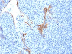
Biotium Cytokeratin 16 (KRT16) (Suprabasal Keratinocyte Marker)(LL025), CF740 conjugate, 0.1mg/mL
Cytokeratins are a family of intermediate filament proteins that assemble into filaments through forming heterodimers of one type I Cytokeratins (Cytokeratins 9 to 23) and one type II Cytokeratins (keratins 1 to 8). The cytokeratin proteins play a critical role in differentiation, as well as tissue specialization and function, to maintain the overall structural integrity of epithelial cells. Cytokeratins are also useful markers in identifying the origin of metastatic tumors. Cytokeratin 16 is expressed in benign stratified squamous epithelium and squamous cell carcinoma of the head and neck, as well as luminal cells of mammary gland and sweat ducts. It is absent in non-invasive breast carcinomas and normal breast tissue. Primary antibodies are available purified, or with a selection of fluorescent CF Dyes and other labels. CF Dyes offer exceptional brightness and photostability. Note: Conjugates of blue fluorescent dyes like CF405S and CF405M are not recommended for dete
Biotium MAML3 (Mastermind Like Transcriptional Coactivator 3) (MAML3/1303), CF568 conjugate, 0.1mg/mL
MAML3 (mastermind-like protein 3) is a nuclear speckle protein that acts as a transcriptional coactivator for Notch receptors. The Notch signaling pathway influences cell fate by regulating the ability of precursor cells to properly respond to developmental signals. MAML3 is a member of the mastermind-like family of proteins that are human homologs of the Drosophila melanogaster mastermind protein. Through its N-terminal region, MAML3 interacts with the ankyrin repeats of the Notch proteins Notch 1, Notch 2, Notch 3 and Notch 4.This interaction leads to formation of a DNA-binding complex with the Notch proteins and RBP-J, a complex that can then induce HES1 gene expression. While the N-terminal domain of MAML3 is essential for proper Notch binding, the C-terminal domain of MAML3 is essential for transcriptional activation. Due to its involvement in cell signaling and transcriptional activation, upregulation of MAML3 is thought to be involved in oncogenesis.Primary antibodies
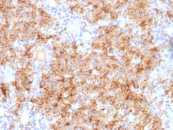
Biotium Chromogranin A / CHGA (Neuroendocrine Marker), 0.2mg/mL
Chromogranin A is present in neuroendocrine cells throughout the body, including the neuroendocrine cells of the large and small intestine, adrenal medulla and pancreatic islets. It is an excellent marker for carcinoid tumors, pheochromocytomas, paragangliomas, and other neuroendocrine tumors. Co-expression of chromogranin A and neuron specific enolase (NSE) is common in neuroendocrine neoplasms. Reportedly, co-expression of certain keratins and chromogranin indicates neuroendocrine lineage. The presence of strong anti-chromogranin staining and absence of anti-keratin staining should raise the possibility of paraganglioma. The co-expression of chromogranin and NSE is typical of neuroendocrine neoplasms. Most pituitary adenomas and prolactinomas readily express chromogranin. Primary antibodies are available purified, or with a selection of fluorescent CF Dyes and other labels. CF Dyes offer exceptional brightness and photostability. Note: Conjugates of blue fluorescent dyes